Dendritic Spines
Dendritic spines are a common feature of post-synaptic processes in excitatory synapses of the Central Nervous Sysytem. Their putative function will be examined in this exercise
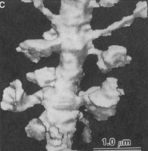
Reconstruction of a CA1 pyramidal cell dendrite
Lisman and Harris, 1993
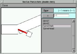
A spine is modeled here as two abutting cylinders with the spine head having a length and diameter of 0.5 microns and the spine stalk having a length of 1.0 microns and diameter if 0.1 microns. This structure is connected to a dendrite 1.0 microns in diameter and 4,000 microns in length.
Bring up the simulation by typing: spike spine.hoc -
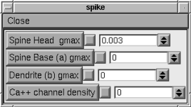
We will be examining the voltage excursions to alpha-function conductance changes elicited in both the spine head and the spine base.
1. Passive spread
After choosing ‘Keep Lines’‘ in both graph windows. Stimulate first in the Spine Head (as shown) and then in the Spine Base with 0.003 uS. What general statements can you make about current spread.with this geometry?
2. Calcium channels
Erase lines in both graph windows. Using a 0.003 uS.stimulus in the spine head only, run the simulation. Now increase the ‘Calcium Channel Density’ from 0 to 2.2 S./cm2 and run the simulation again. Observe the effect of the Calcium channel.
3. Effects of Geometry
Change the length and diameter of the Spine Stalk. Make some general observations of this manipulation.
4. A third stimulation point is positioned 200 microns away from the spine base on the dendrite (‘Dendrite (b)’ on the control panel). Use a 0.003 uS gmax with and without Calcium channels and observe the voltage at the Spine Head and Base.